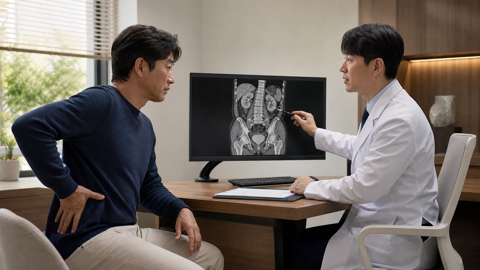
Extracorporeal shock wave lithotripsy is a treatment that focuses shock waves from outside the body on kidney stones to break them into small pieces, allowing the fragments to be expelled through urine. Although it seems less burdensome due to the advantage of no skin incisions or endoscope insertion, not all stones break perfectly at once and disappear immediately.
The results are influenced not only by the size of the stone but also by its location and hardness, the distance from the skin to the stone, the ability to target it, and the pathway through which urine can pass. Therefore, you should first check whether ‘my stone is in a condition that can break easily and pass well’ before considering the procedure time.
Focus the shockwave on the point of absence
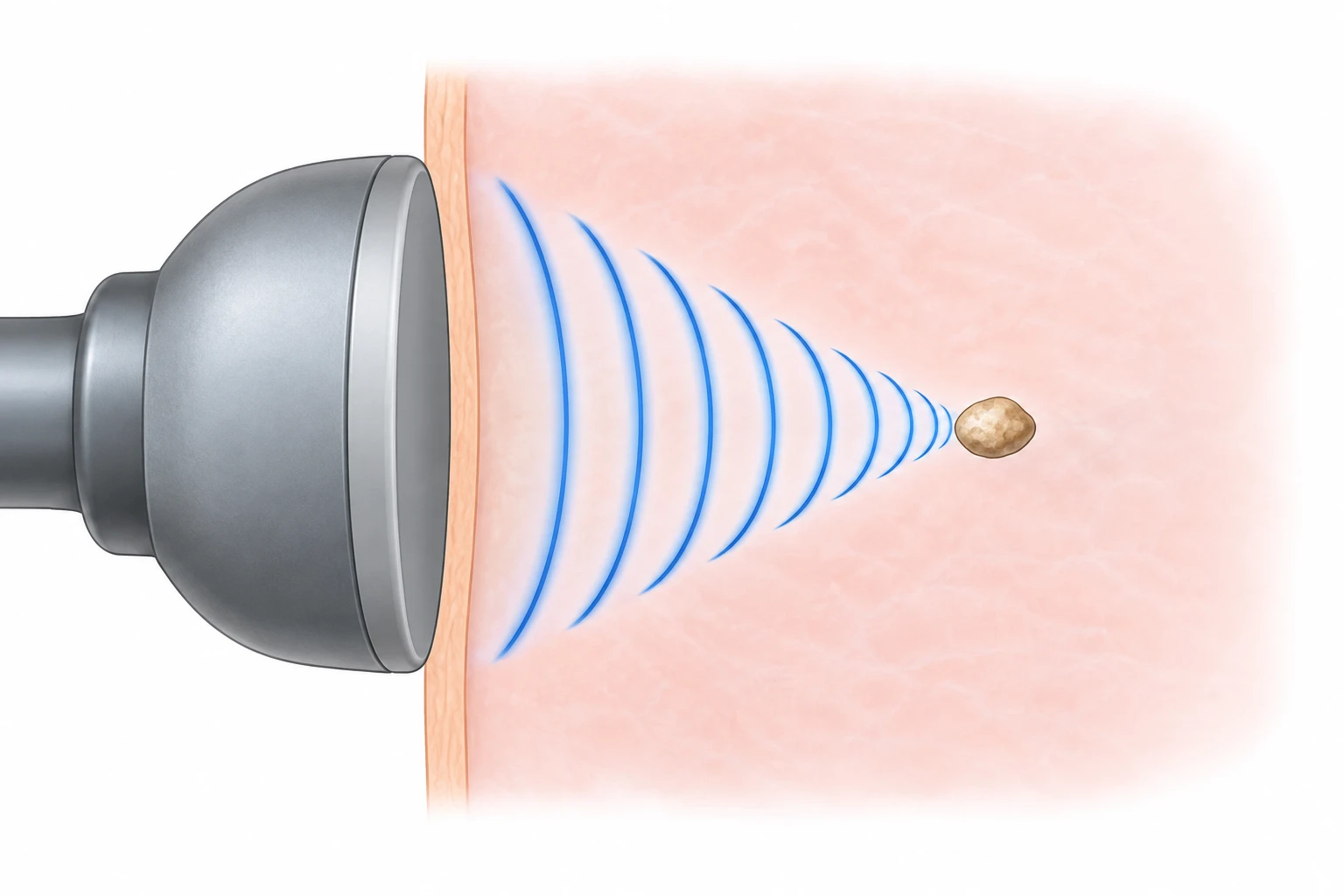
When lying on the treatment table, the stone’s location is identified using X-ray or ultrasound, and shock waves are delivered from the treatment head in contact with the skin. The energy starts low and is gradually increased, while it is checked during treatment that the stone does not move out of the focus. The EAU guidelines emphasize that precise targeting, imaging monitoring, and appropriate pain management are important for the outcome.
Movement or irregular breathing caused by pain can make it harder to keep the shock waves accurately focused. Rather than trying to endure significant discomfort, tell the medical team so they can adjust pain control or the treatment settings. Anesthesia and sedation practices vary by equipment, facility, and patient condition, so confirm fasting requirements and whether you will need someone to accompany you home when scheduling the procedure.
Size, location, and CT density determine the likelihood of success
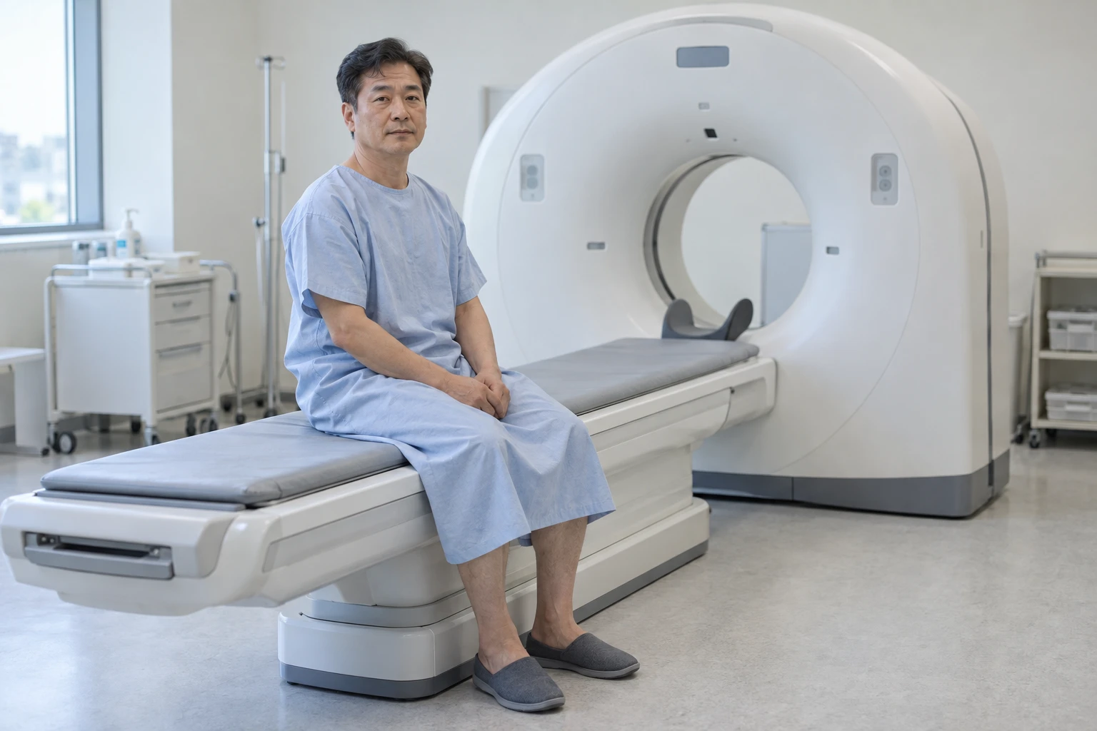
Relatively small stones in the upper or middle calyces of the kidney, which are well visualized and easy to target on imaging, can be considered for extracorporeal shock wave lithotripsy. On the other hand, stones in the lower part of the kidney may be difficult to pass even if broken due to anatomical angles and gravity. The larger the stone, the higher the possibility of requiring multiple treatments.
If the Hounsfield units on a non-contrast CT are high, making the stone appear hard, or if components that are difficult to break with shock waves, such as calcium oxalate monohydrate, brushite, or cystine, are expected, the chances of success may decrease. If the distance from the skin to the stone is long or there is an anatomical obstruction in the distal part, other methods may be more suitable. Compare the possibility of stone removal, invasiveness, and the potential for retreatment together between ureteroscopic or percutaneous nephrolithotomy.
Check the risk of infection and bleeding the day before
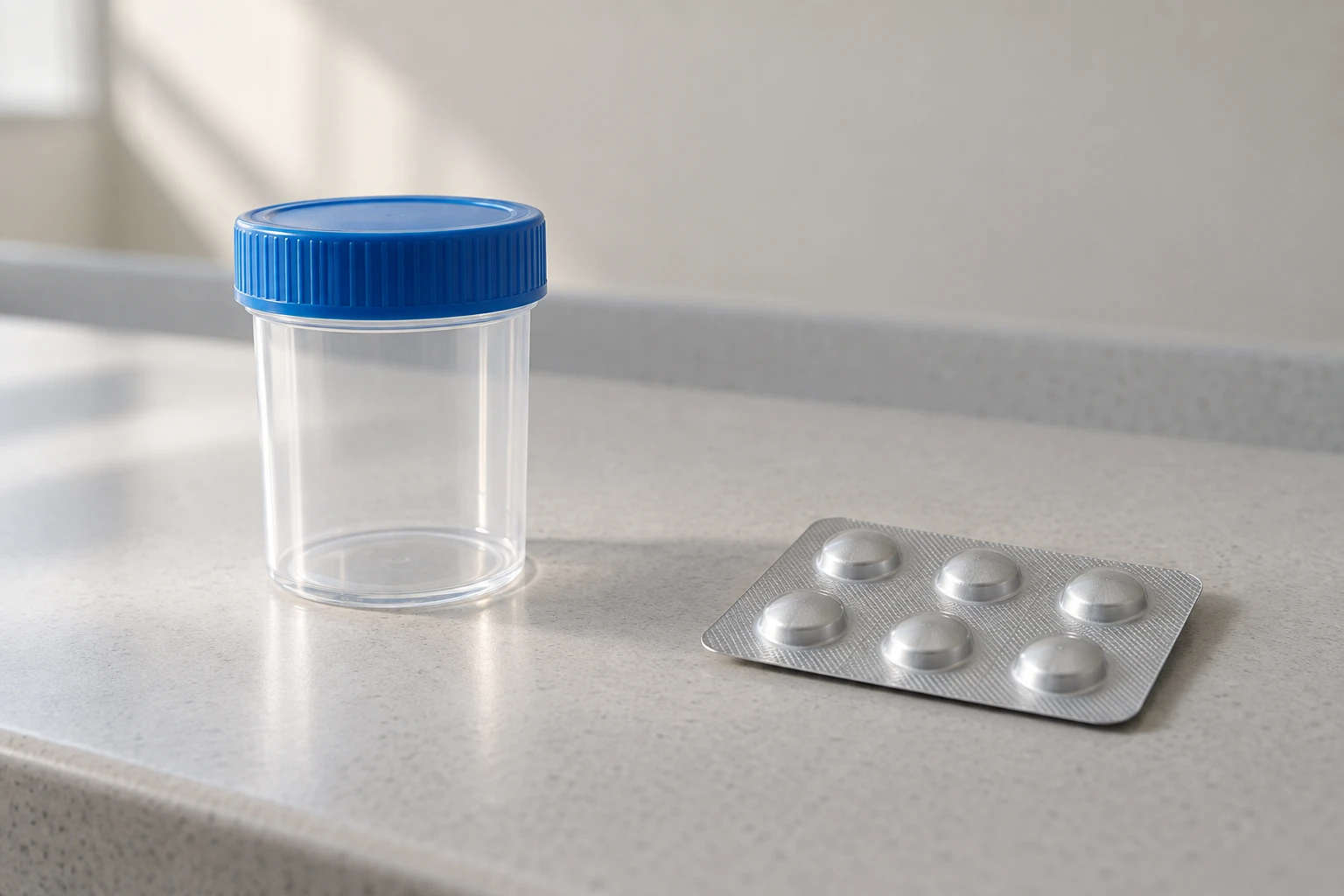
Before the procedure, a urine test is performed to check for infection. If an infected urine is blocked by a stone causing fever and chills, it may be necessary to secure the urinary tract and treat the infection before lithotripsy. Uncontrolled urinary tract infection, pregnancy, uncorrected bleeding disorders, aneurysms around the stone, conditions that make targeting difficult, and obstruction below the stone may be contraindications or unsuitable factors for extracorporeal shock wave lithotripsy.
Aspirin, clopidogrel, warfarin, and direct oral anticoagulants affect the risk of bleeding. However, stopping them arbitrarily can increase the risk of stroke or thrombosis, so you should accurately inform your doctor of the medication names and reasons for taking them and receive adjustments. Also, inform them in advance about potential pregnancy, implantable devices such as pacemakers, and previous stone analysis results.
Stone fragments move after the procedure
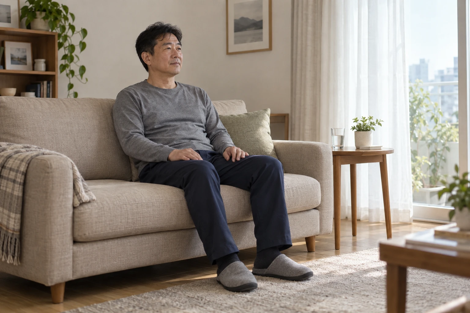
After the procedure, you may experience mild hematuria, flank discomfort, and light bruising on the skin. As stone fragments pass down the ureter, colic may occur again, so check the prescribed pain medication and emergency contact methods. If you do not have heart or kidney disease that requires a fluid restriction, you can drink fluids within the recommended range, and you can collect the expelled fragments using a strainer for component analysis.
A ‘stone street’ (steinstrasse), where multiple fragments line up in the ureter, can block the flow of urine. High fever around 38 degrees, chills, inability to urinate, pain that cannot be controlled with analgesics and repeated vomiting, a large amount of blood in the urine, or large clots are signs that require immediate medical attention without waiting.
Follow-up imaging is necessary even if the pain disappears

The disappearance of pain does not mean that the stone has been completely expelled. At the designated time, remaining stones and obstruction should be checked using X-ray, ultrasound, or CT. Depending on the location and size of the remaining stones, the next steps, such as observation, repeat lithotripsy, or ureteroscopy, may vary.
After removing kidney stones, it is also necessary to examine the cause of recurrence. Even if it is the first stone, its composition should be analyzed if possible, and for recurrent stones, bilateral stones, early-onset cases, or in the presence of certain diseases, blood tests and 24-hour urine tests should be considered. Rather than just drinking a lot of water, a prevention plan tailored to the stone composition, diet, and underlying conditions is important.
Extracorporeal shock wave lithotripsy is not a treatment chosen with the single reason of having no incision. The conditions of the stones observed in imaging, the possibility of repeated treatments, stone fragment passage, and follow-up examinations should all be understood as one treatment process.
Referenced Medical Information
- European Association of Urology (EAU) 2026 Urolithiasis Guidelines
- European Association of Urology (EAU) 2026 Changes Summary
This article is intended to provide general health information and does not replace diagnosis or treatment. The treatment method for stones should be determined with medical professionals based on their location, size, composition, infection, and overall health condition.