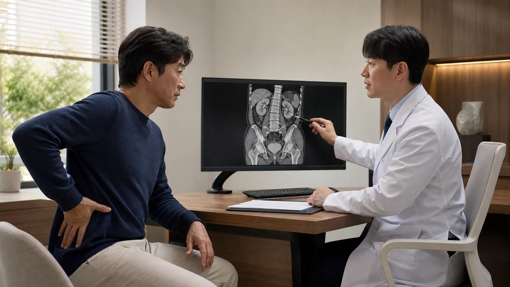
Extracorporeal shock wave lithotripsy is a treatment that breaks stones without opening the body. More precisely, it involves concentrating shock waves generated outside the body on a single point of the stone and hitting it repeatedly. When researching, it is often confused with extracorporeal shock wave therapy (ESWT) used in orthopedics, but the treatment for musculoskeletal pain and extracorporeal shock wave lithotripsy (ESWL) for urinary stones are completely different procedures that only share a similar name. This article does not compare specific hospitals. It will go over the principle of the procedure, the suitable conditions, pain and cost, and its limitations in order.
Shock waves converge on a stone at a single point and create cracks

The key to extracorporeal shock wave lithotripsy is the ‘focus.’ It is a non-invasive method designed to concentrate high-energy acoustic shock waves generated outside the body onto a specific point where the stone is located, while allowing the energy to dissipate in tissues beyond that point. Depending on the method used to generate the shock waves, the equipment is classified into electrohydraulic, electromagnetic, and piezoelectric types, and in the electromagnetic type, a protocol that starts at a low voltage and gradually increases is commonly used.
So how does the wave of sound break a stone? When a shock wave passes through body fluids, it creates a sudden change in pressure in that spot. At this time, the phenomenon of cavitation, where tiny bubbles are formed and then collapse, occurs, and the energy released from this, along with the repeated tensile and compressive stresses accumulating on the surface and inside the stone, causes cracks in the stone. It is not broken in a single shot; rather, it is more like the principle of repeatedly tapping the same spot thousands of times to grow a crack.
The number of shock waves applied per second also changes the results. Studies continue to address the fact that adjusting the frequency affects both the stone removal rate and the extent of surrounding tissue damage, and standards vary depending on the equipment and institution. Even for procedures with the same name, the detailed protocols have not yet been finalized, so asking in the clinic, ‘What method does this equipment use and how many sessions are planned?’ is by no means an unreasonable question.
Once you understand the principle, the difference from other treatments becomes clear. Ureteroscopic stone removal (URS) involves inserting an endoscope directly through the urethra into the ureter and kidney to visually locate the stone, then breaking it with a laser upon contact. Whether or not the stone is touched, this point of contact is what distinguishes the principles of the two treatments, and most of the differences in success rates and recovery discussed later also stem from this.
Determines whether the size, density, and location are appropriate
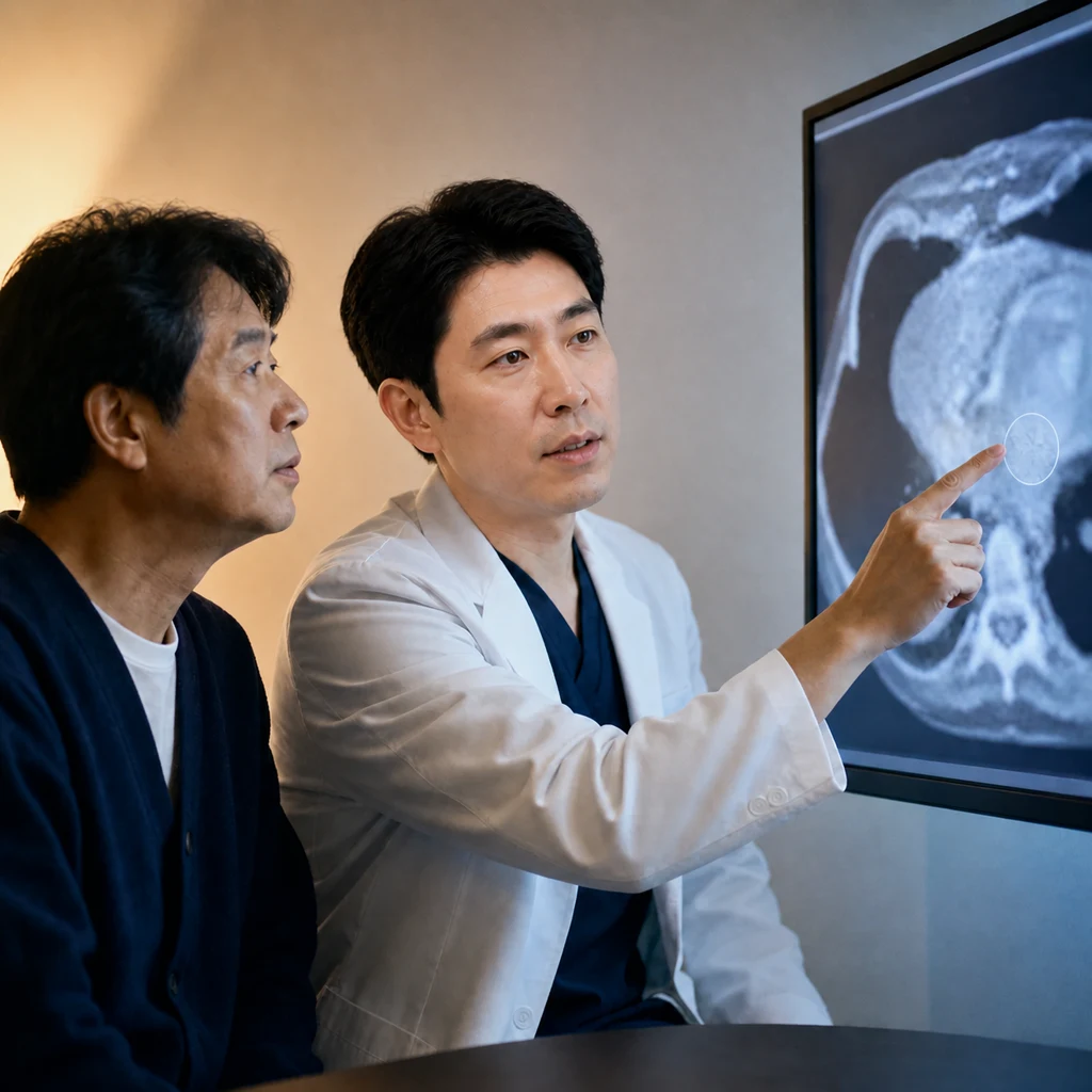
Extracorporeal shock wave lithotripsy is not a procedure that yields the same results for everyone. Three factors—the size, density, and location of the stone—greatly influence its suitability. The primary recommended candidates are kidney stones smaller than 2 cm and upper ureter stones.
Above all, the harder the stone, the less easily it breaks. The lower the stone density measured on a CT scan, the more easily it can be crushed, whereas dense, hard stones do not crack easily even when the same energy is applied repeatedly. This is exactly why the composition and density of the stone are examined in advance through imaging before the procedure.
Location also affects outcomes. Multiple studies have repeatedly reported that stones in the upper ureter have better results than stones in the middle or lower ureter, but since the definition and figures for success rates vary across studies, there is no need to accept a specific percentage as definitive. Stones located in the lower part of the kidney (lower pole) are structured in a way that, even if fragmented, they must move against gravity to be expelled, so the European Association of Urology recommends considering stones up to 20mm for treatment only if there are no unfavorable factors.
There are also conditions that are difficult to implement in the first place.
- If pregnant
- In cases with bleeding tendency not corrected by medication
- In cases of uncontrolled urinary tract infection
- In cases of severe obesity or significant skeletal deformity, it may be difficult for the shockwave focus to properly reach the stone
- Lower pole urinary tract obstruction when there is no path for the fragmented pieces to pass
- If there is an aneurysm near the missing area
This is the reason why the approach of ‘if it’s a urinary stone, start with lithotripsy’ does not hold. Pre-procedural imaging is not just a formal procedure, but a key basis for determining suitability.
Pain during the procedure and pain during discharge are different events
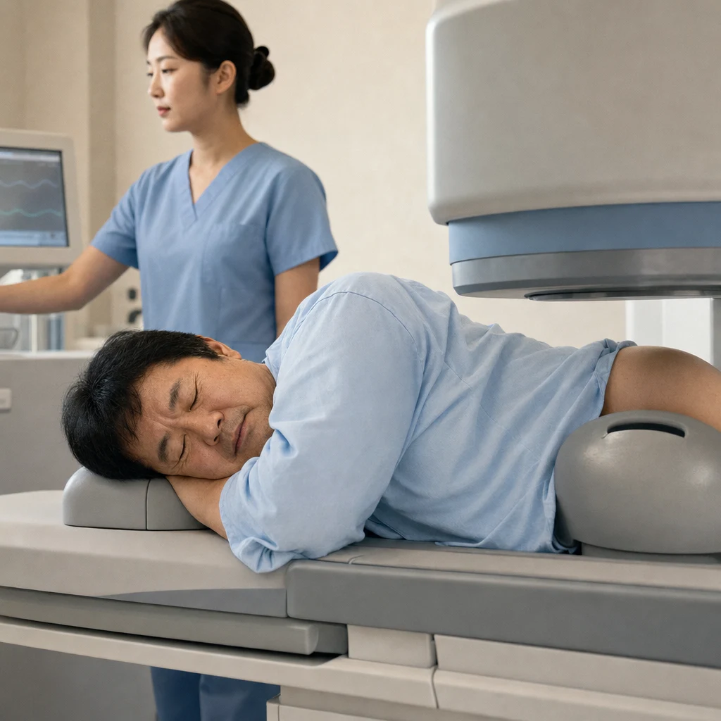
The duration of a single procedure is generally guided to be in the range of approximately 30 to 60 minutes. Most procedures are performed on an outpatient basis without hospitalization, and anesthesia is generally not used, with pain being managed with analgesics if necessary. One point of comparison with surgical treatment is that in many cases, patients return to their daily activities on the same day immediately after the procedure.
The questions that come up most frequently in the consultation room are concentrated in this area. They are about how painful the procedure is and whether anesthesia is used. The sensation of shockwaves passing through the body is often described as tingling or a stiffness that feels like being snapped by a rubber band, and there is a large individual difference in the level of sensation even under the same conditions. The fact that it is difficult to agree on the pain level with a number in advance is itself a recurring characteristic of this procedure.
The side that actually requires mental preparation is after the procedure. During the several days to weeks while fragments of the broken stone travel down the ureter, discomfort such as flank pain and blood in the urine may follow. This pain is a separate event from the pain of the procedure itself, and if you do not distinguish between the two, misunderstandings like ‘They said it wouldn’t hurt, so why does it hurt so much?’ can arise. The procedure is over, but what happens inside the body really starts from that point.
It is better to know about the side effects along with their frequency of occurrence.
- Pain during and after the procedure, labor-like pain, hematuria, fever: These are relatively commonly reported reactions.
- Ureteral obstruction by fragmented stones (steinstrasse): This is a condition where fragments line up and block the ureter, with reported incidence ranging widely from 1.1% to 24.2%. For stones larger than 2 cm, it is reported in 5–10% of cases, and for partial staghorn stones, up to 40%, with the risk increasing as the stone size increases. Additional treatment is required if colic or infection occurs.
- Perirenal hematoma: Asymptomatic hematomas are observed in about 15% of cases and cannot be considered a rare complication. Cases accompanied by symptoms are relatively few, about 1%. Hypertension, coagulopathy, history of previous lithotripsy, and large stones are considered risk factors.
Therefore, if you are taking blood pressure medication or anticoagulants, or have previously undergone lithotripsy, you must disclose this during the pre-procedure consultation. Controlling blood pressure and managing anticoagulants are practical measures to reduce the risk of hematoma.
Lithotripsy for urinary stones is a reimbursable item on an outpatient basis

Extracorporeal shock wave lithotripsy for urinary stones is a procedure covered by health insurance on an outpatient basis. This point is precisely where it differs from shock wave therapy in orthopedics. If the per-session cost information of non-covered extracorporeal shock wave therapy used for musculoskeletal purposes is directly applied to urinary stone lithotripsy, the expected financial burden will differ significantly.
Because it is covered by Korea’s National Health Insurance, the cost burden is generally lower than paying the full amount for a non-covered service. However, the actual out-of-pocket expense varies by hospital type and individual insurance status. Publicly available estimates span a wide range, so it is difficult to provide a specific amount in advance. The most reliable approach is to ask the treating medical facility for a current estimate.
There are items that are often omitted in cost calculations. Imaging tests such as CT or ultrasound before the procedure are billed separately from the cost of lithotripsy, and if the stones are not sufficiently broken and a secondary treatment such as ureteroscopy is needed, that cost occurs as a completely separate item. When estimating the total cost, it is better to check these two in advance.
If it doesn’t finish in one go, the ureteroscope is next
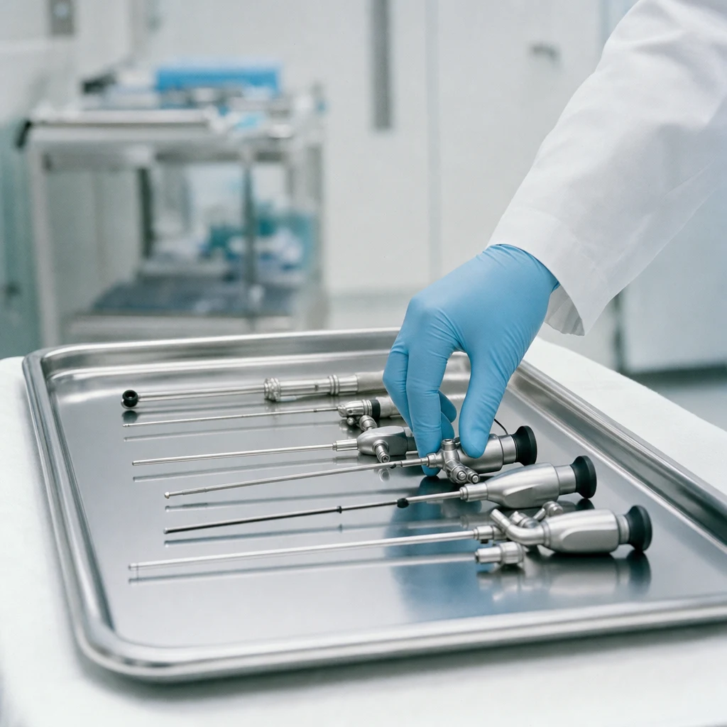
Extracorporeal shock wave lithotripsy is not the only solution for treating urinary stones, but rather occupies one step in a staged treatment structure. The advantages of not inserting instruments into the body are particularly highlighted when the stones are small, low in density, and located in the upper ureter or kidney. Conversely, if the stones are large, located in the lower calyx, or have a history of not being sufficiently broken with a single procedure, ureteroscopic stone removal (URS) or percutaneous nephrolithotomy (PCNL) are considered as alternative options.
Ureteroscopy involves inserting an endoscope through the urethra under anesthesia to directly access the stone, then breaking it with a laser and removing the fragments. The practical difference from extracorporeal shock wave lithotripsy is that ureteroscopy requires more anesthesia and hospitalization. On the other hand, it allows direct visualization of the stone while handling it and immediate retrieval of the fragments. The pattern of complications is also different: lithotripsy more commonly causes pain, renal colic, and fever, while ureteroscopy more frequently results in hematuria and urosepsis after the procedure.
Attempts to determine superiority between two treatments solely by success rate are not as appropriate as one might think. One direct comparison study reported stone removal rates of 49.2% for extracorporeal shock wave lithotripsy and 57.8% for ureteroscopy. Meanwhile, another study examining only shock wave lithotripsy reported stones up to 77~82% when limited to stone size and location. These two figures differ in study design and the conditions for the target stones, so they cannot be compared on the same scale. Simply moving numbers without prerequisites can easily cause misunderstandings.
Therefore, it is risky to oversimplify by saying ‘this is definitely better because it’s not surgery.’ The medical judgment that a procedure is less invasive and suitable for my stone are entirely different issues. If the stone does not break completely in one session, repeated procedures are possible, and if fragments remain after repetition, switching to endoscopic treatment is a common stepwise approach. Keeping this overall structure in mind in advance reduces the chances of being caught off guard when the next steps are discussed.
You need to start with the possibility of recurrence in mind

Extracorporeal shock wave lithotripsy is a procedure where the possibility of repeat treatment remains open depending on the characteristics of the stones. Although there are comparative studies suggesting a higher repeat procedure rate than ureteroscopy, these are only single studies and the variation according to stone conditions is large. Therefore, it is practically more important to recognize in advance the possibility that a single procedure may not be sufficient, rather than focusing on specific numbers.
It is also a common misconception that ‘once you receive it, the stones completely disappear and never come back.’ Even after the procedure, follow-up imaging is necessary to check for any remaining stones, and since stones are a condition that frequently recurs, consistent management, including fluid intake, must continue. Setting the date for the procedure is not the end of management but a new beginning.
Before deciding on the procedure, please check the following three things with your doctor in charge.
- Are the size, density, and location of my defect suitable for this procedure? I need to hear an explanation based on the results of imaging tests.
- To what extent is repeated treatment possible, and after how many attempts is the next step considered?
- During the discharge process, what is within the expected range, and what symptoms require an immediate visit to the hospital?
If you start by clearly knowing these three answers, you can naturally accept the pain for a few days after the procedure as a planned process. A significant part of the preparation needed for stone treatment is determined right here.