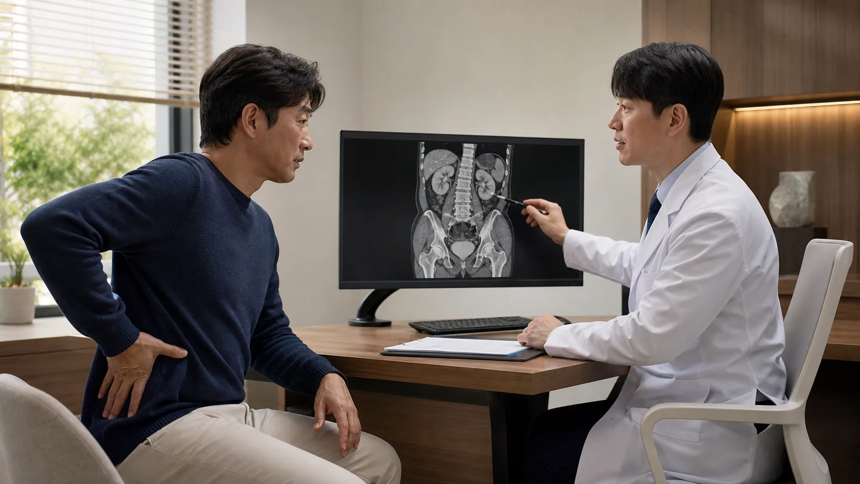
The moment you search for ‘Gangnam urinary stones’ usually comes without warning. Pain that starts on one side of the flank can spread to the lower abdomen or groin, and if changing positions doesn’t bring relief, you might think of stones. However, you cannot determine the size or risk of the stone solely based on the intensity of the pain.
Urolithiasis is a condition in which crystals clump together to form stones in the paths through which urine passes, such as the kidneys, ureters, and bladder. Currently, it is necessary to both reduce pain and check for infection or obstruction, and then decide whether to wait for natural excretion or actively remove the stones.
If fever and chills accompany, it is not considered simple labor pain
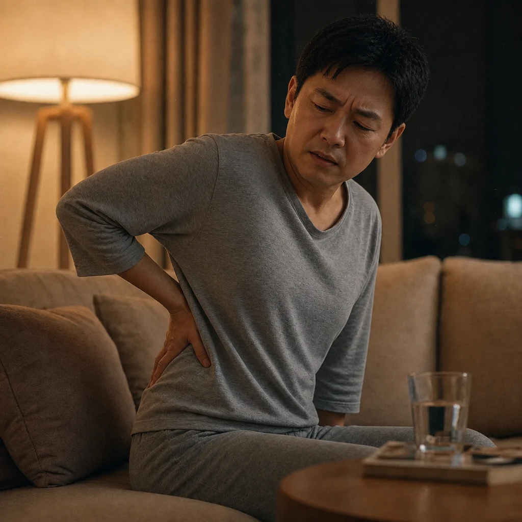
The pain from kidney stones often worsens and eases like waves, and may be accompanied by nausea, vomiting, and blood in the urine. On the other hand, if there is fever and chills, very little urine output, pain that cannot be controlled with painkillers, or repeated vomiting, prompt evaluation is necessary.
In particular, if infected urine is blocked below a stone, it can become a urological emergency. In this case, it may be prioritized to first drain the blocked urinary tract and treat the infection rather than immediately breaking the stone. Obstruction in both ureters or in a person with only one kidney should also be urgently evaluated to protect kidney function.
Urine tests and imaging tests answer different questions
A urine test checks for hematuria and signs of inflammation, and if necessary, a culture test is used to identify infectious bacteria. A blood test is used to examine kidney function, inflammation, and metabolic items such as calcium and uric acid.
Ultrasound can detect kidney swelling and some stones without radiation exposure. Non-contrast CT provides detailed information on the location, size, density of stones, and the degree of obstruction, helping to guide treatment choices. Not everyone needs the same tests, and the order of examinations is determined considering the possibility of pregnancy, age, symptoms, and previous imaging results.
Waiting, shockwave, and endoscopic treatment are selected according to conditions
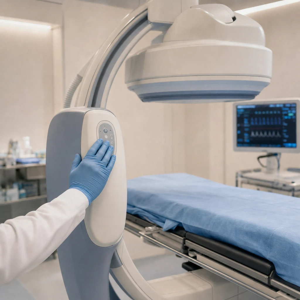
Small ureteral stones without complications can be monitored for natural passage while managing pain. However, you should consider not only the small size but also pain, infection, kidney function, and stone location rather than waiting for a long time. For some ureteral stones, medications that help with passage can be considered, but they do not provide the same benefit for all locations and sizes.
Extracorporeal shock wave lithotripsy is a method that focuses shock waves from outside the body to break kidney stones into small pieces. It has the advantage of no incisions, but depending on the location, size, and hardness of the stones and the distance from the skin to the stones, repeated procedures or additional treatment may be necessary.
Ureteroscopy involves inserting an endoscope through the urethra to directly crush or remove stones. While there is a high possibility of stone removal, factors such as anesthesia, ureteral stents, and discomfort after the procedure are also considered. For large kidney stones or complex stones, percutaneous nephrolithotomy, which creates a small passage in the back, may be more suitable.
Prevention of recurrence begins after an absence occurs

Stone fragments that are expelled or removed are analyzed for their composition whenever possible. This is because the prevention strategy varies depending on the type of stone, such as calcium stones, uric acid stones, or infection stones. In cases of recurrent stones, early-onset, bilateral stones, or a family history, metabolic evaluation such as a 24-hour urine test may be considered.
The starting point for general prevention is to drink water throughout the day to ensure sufficient urine output. It is also important to reduce salt intake, have a balanced diet including vegetables and fruits, and avoid consuming excessive amounts of animal protein. Arbitrarily and excessively restricting dietary calcium due to calcium stones can actually increase oxalate absorption in the intestines, so adjustments should be made according to individual results.
In Gangnam urinary stone treatment, you need to plan both ‘how to deal with the stone that is causing pain now’ and ‘how to reduce the next stone.’ Even after the pain disappears, make sure to check whether follow-up imaging and evaluation of composition and metabolism are necessary.
Referenced Medical Information
- European Association of Urology (EAU) 2026 Urolithiasis Guidelines
- European Association of Urology (EAU) 2026 Changes Summary
This article is intended to provide general health information and does not replace diagnosis or treatment. If you experience fever, chills, decreased urine, or uncontrolled pain, seek prompt evaluation by a medical facility.