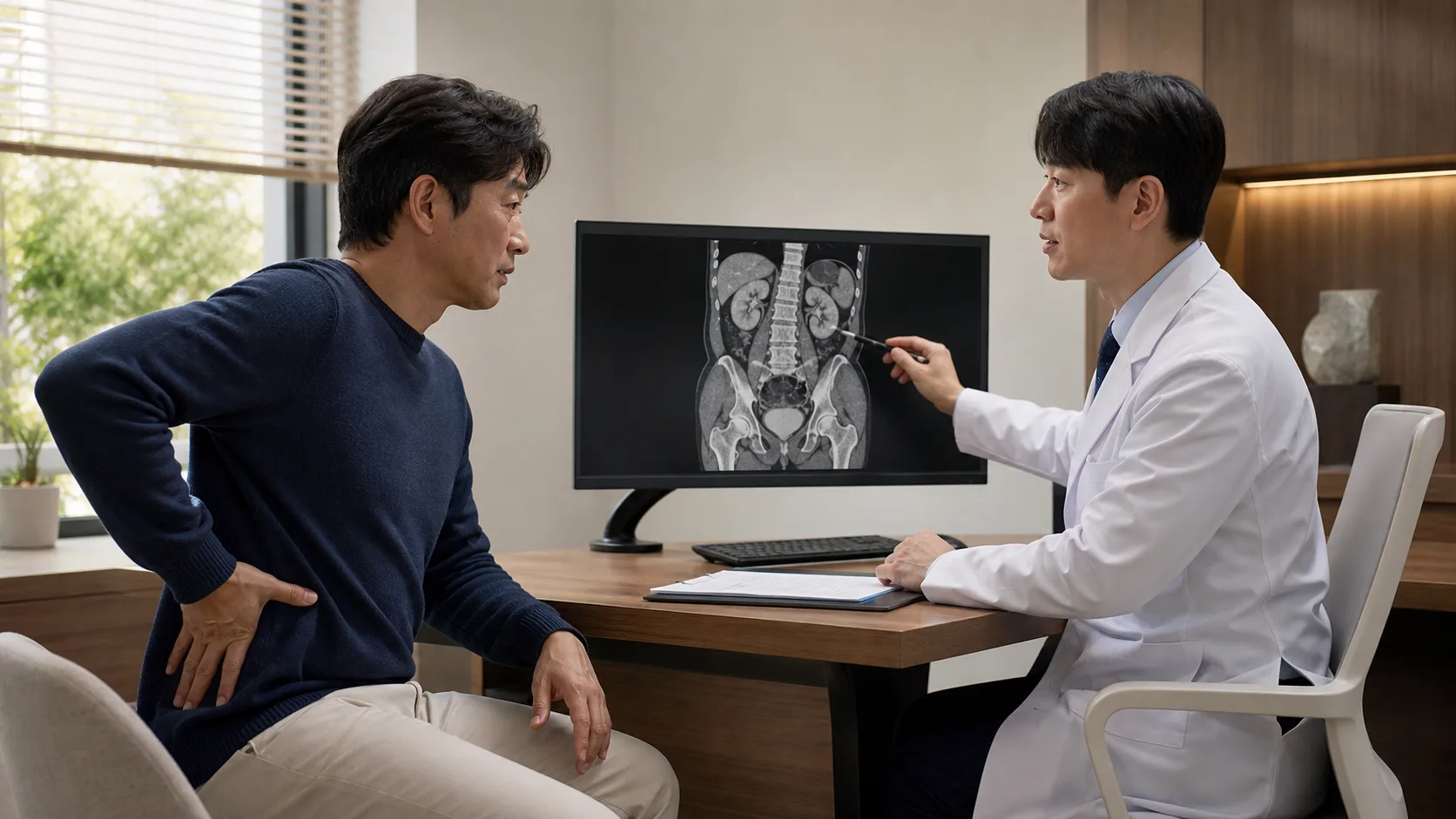
If you search for ‘Gangnam Extracorporeal Shock Wave Lithotripsy,’ the first explanation you will see is that it ‘breaks stones without an incision.’ While this is correct, it does not mean that the same result is guaranteed for every stone. The outcome depends on where the stone is located, how large and hard it is, whether it can be accurately targeted from outside the body, and whether there is a path for the broken pieces to pass.
Extracorporeal shock wave lithotripsy (SWL or ESWL) has the advantage of being a less invasive option, but it is important to understand the possibilities of repeated treatments and residual stones. Compared to endoscopic treatment, which option to prioritize depends on individual conditions.
Shock waves gather energy on kidney stones from outside the body
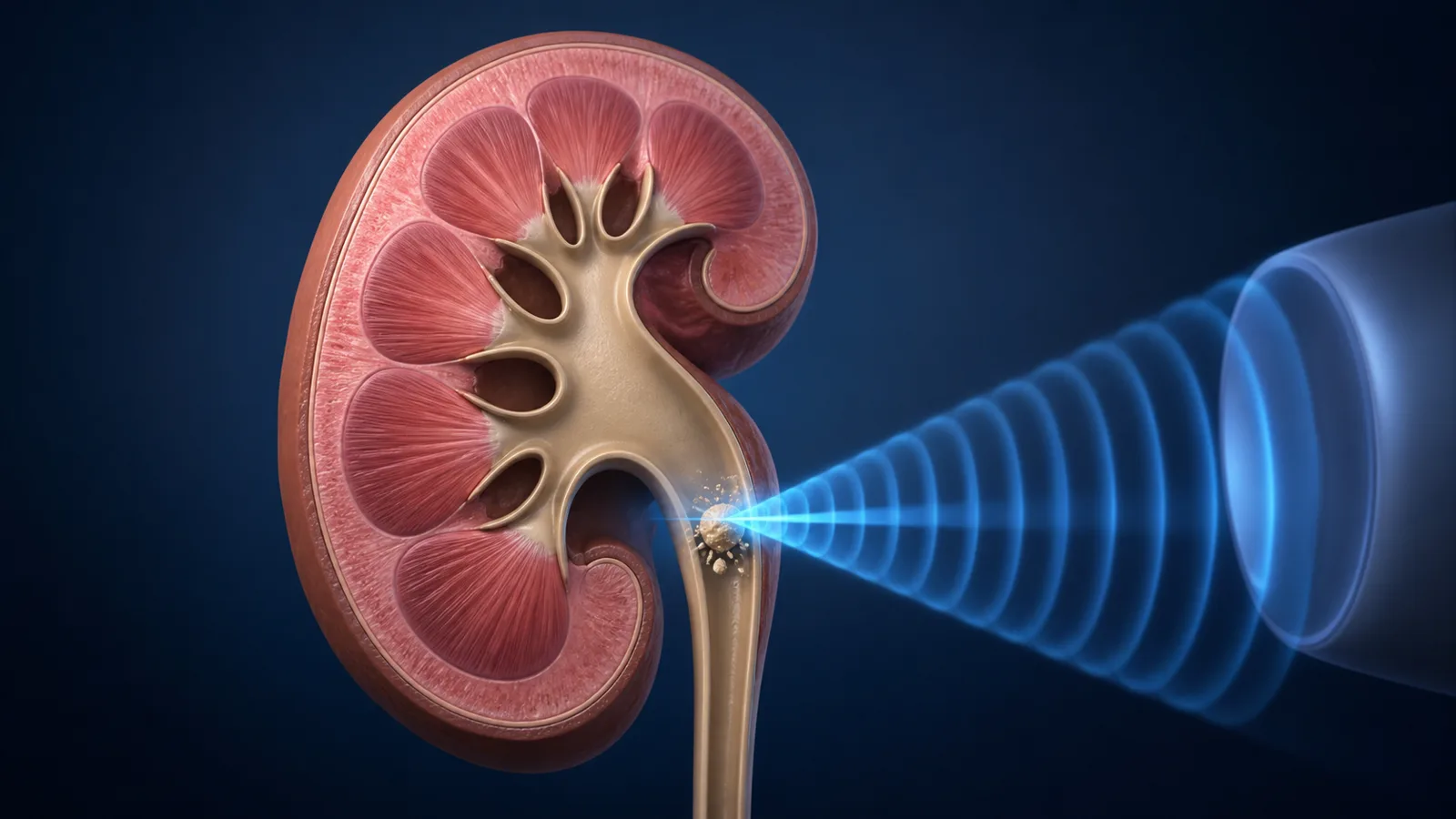
While lying on the treatment table, X-rays or ultrasound are used to locate the stone, and shockwaves are delivered through a treatment head in contact with the skin. The energy repeatedly concentrates on the stone, breaking it into small pieces, which are then expelled through urine. Since this is not a surgery to directly remove the stone, time is needed for the fragments to be expelled.
Accurate targeting, the coupling between the skin and the treatment head, and proper pain control affect the results. If the body keeps moving due to breathing or pain, the focus can become blurred, so it is important to maintain your posture according to guidance during the procedure.
Size, location, and firmness change the chances of success
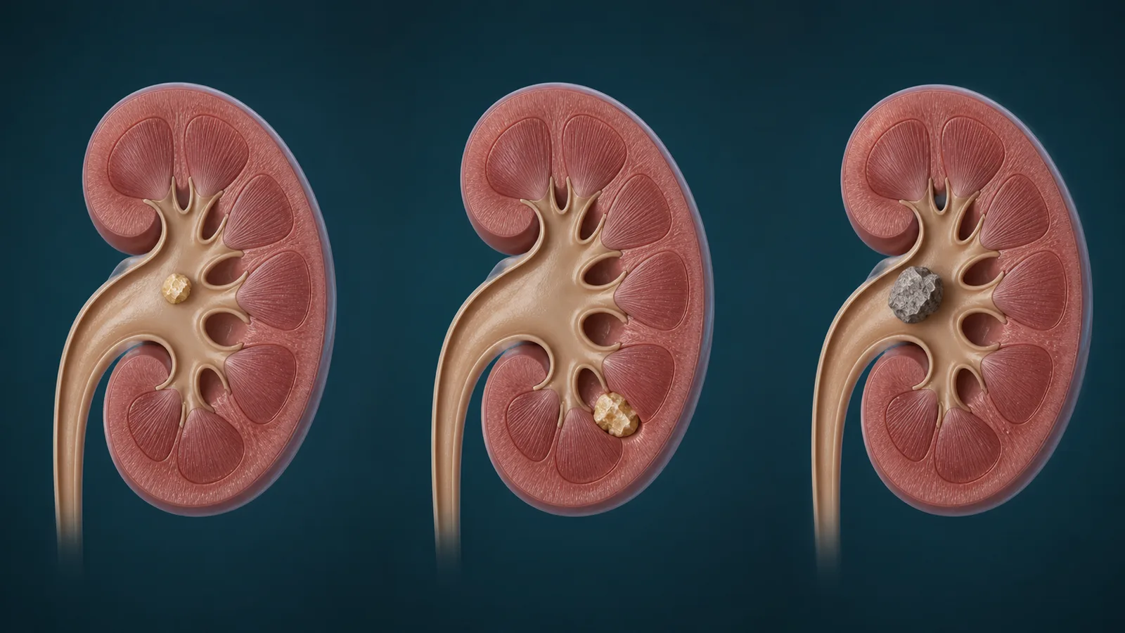
The 2026 EAU guidelines explain that SWL, ureteroscopy, and percutaneous nephrolithotomy should be selected according to the size and conditions of stones in the renal pelvis and upper/mid calyces. In general, the larger the stone, the lower the stone clearance rate with SWL and the higher the likelihood of needing repeated treatment. Lower calyx stones may fragment well, but due to gravity and anatomical structure, it can be difficult to expel the fragments.
Stones with high density on CT, such as calcium oxalate monohydrate, brushite, and cystine, which are hard components, may not break easily with shock waves. The results can also be affected if the distance from the skin to the stone is long or if aiming is difficult due to the skeleton or body shape. This is why, in consultations for extracorporeal shock wave lithotripsy in Gangnam, they ask not only about the size in millimeters, but also about the location, CT density, the expected composition, and the passage for discharge.
First, check the risk of infection and bleeding, and contraindications
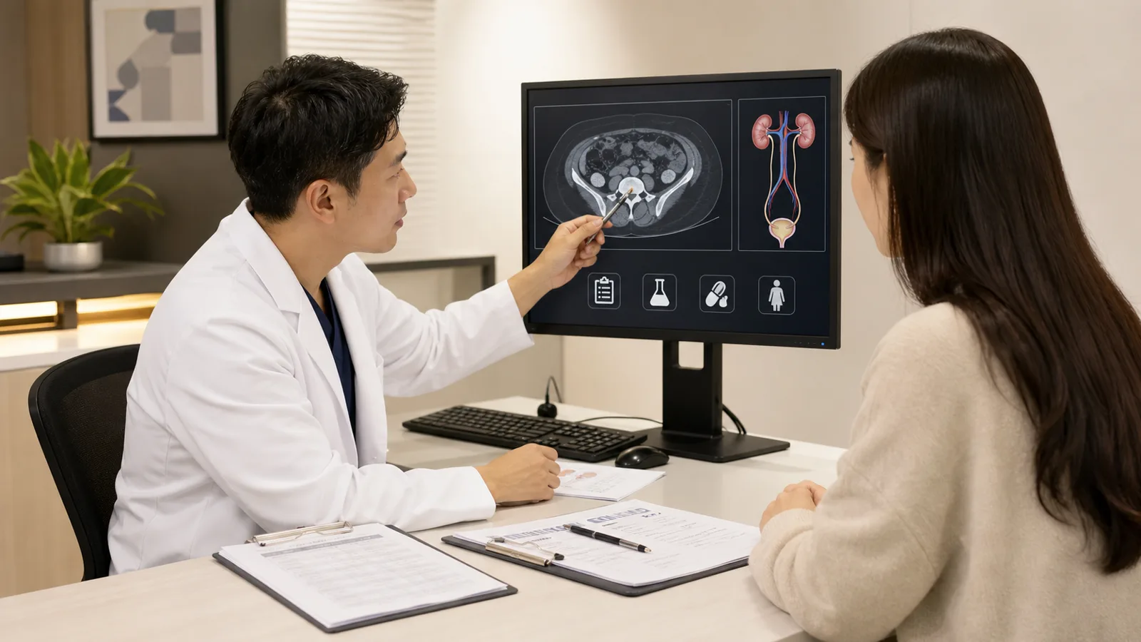
Pregnancy, uncorrected bleeding disorders, uncontrolled urinary tract infections, and anatomical obstruction below the stone are important contraindications for extracorporeal shock wave lithotripsy. Aneurysms around the stone, severe skeletal deformities, or obesity that makes targeting difficult can also affect the treatment plan.
Urinary tract infection is confirmed through a urine test and, if necessary, a culture test, and it is crucial to inform whether you are taking anticoagulants or antiplatelet drugs. Stopping medication on your own can increase the risk of blood clots, so both the prescribing and procedural medical professionals should plan accordingly. Obstructive stones accompanied by fever and chills are not simply a situation to schedule lithotripsy; it may be necessary to address the infection and blocked urinary tract as an emergency first.
After the procedure, check for stone discharge and emergency signals

After the procedure, a small amount of blood in the urine, flank discomfort, and renal colic caused by passing stone fragments may occur. Adjust your fluid intake and activity according to your kidney and heart condition and the medical staff’s instructions. Collecting the expelled fragments in a clean strainer and storing them can help with stone composition analysis and planning for prevention of recurrence.
Fragments can line up and block the ureter, creating a ‘steinstrasse,’ and in this case, pain, obstruction, and infection can occur. If you experience high fever and chills, severe uncontrolled pain and vomiting, inability to urinate, or significant hematuria or blood clots, you should immediately seek medical attention.
After undergoing extracorporeal shock wave lithotripsy in Gangnam, do not assume that all stones are gone just because the pain has disappeared. It is important to check for residual stones and obstruction through imaging at a designated time, and if it is the first stone or a recurrent case, to continue with component analysis, metabolic evaluation, and fluid and dietary planning.
Referenced Medical Information
- European Association of Urology (EAU) 2026 Urolithiasis Guidelines
- European Association of Urology (EAU) Urolithiasis 2026 Summary of Changes
This article is intended to provide general health information and does not replace diagnosis or treatment. The treatment method for stones should be determined with medical professionals based on their location, size, composition, infection, and overall health condition.